Follow-ups and the progression of rheumatoid arthritis
Published Jun 19, 2019 • Updated Jun 25, 2019 • By Louise Bollecker
Rheumatoid arthritis is a disease that progresses with flare-ups in unpredictable ways. Gradually, if the disease is not treated, it often will affect other joints. This is why it is essential to monitor the progression of the disease in order to control or reduce complications. Follow our explanatory guide.

The first year after the diagnosis of the disease, a monthly assessment (at each consultation) is carried out. When the disease is controlled (remission or low activity) the assessment takes place every 3 months.
Assessing disease progression: the DAS score
DAS - the measurement of disease activity
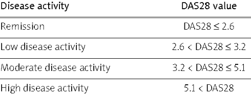
Disease activity is assessed according to certain clinical and biological parameters. These parameters allow the calculation of the DAS 28 score. DAS stands for Disease Activity Score. 28 refers to the joints that are examined in the assessment.
It is an index of rheumatoid arthritis activity that combines several aspects of the disease into a single data set, expressed as a number.

How to calculate the DAS score
The results of the four values below are placed into a mathematical forumla that calculates the overal disease activity score.
1. Swollen Joints
The number of swollen joints (NAG) out of the 28.
2. Tender Joints
The number of tender/painful joints (NAD) out of the 28
3. Patient Global Assessment
The patient assesses his/her global health status.
Using a visual analogue scale from 0 to 10, the patient makes the assessment. 0 = best health / no manifestation of the disease; 10 = worst health / maximum severity of the disease the the patient can imagine.
4. Measure Inflammation: ESR or CRP value
When the body detects substances that seem foreign to it, it sets up a defense strategy to recognize, destroy and eliminate them: this is an inflammatory reaction. The causes of inflammation are multiple: they can be of external origin (bacteria, viruses, skin lesions, etc.) or internal (autoimmune diseases, such as rheumatoid arthritis, cancers...).
- C-Reactive Protein (CRP) is an inflammatory protein, synthesized by the liver, which increases its blood concentration within a few hours during an inflammatory reaction. CRP plays an important role in mobilizing and activating the immune defenses (white blood cells) and stimulating the destruction process of cells considered as foreign (phagocytosis). The higher the CRP value, the more important the inflammatory response.
- Erythrocyte Sedimentation Rate (ESR) is a type of blood test that can help determine the presence of inflammation by measuring how rapidly erythrocytes (red blood cells) settle at the bottom of a test tube containing a blood sample. A medical provider determines the distance erythrocyes fall within a given time (usually one hour). In the presence of inflammation, abnormal proteins (including fibrinogen) are increased, leading to the formation of red blood cell clusters, and thus, causing the erythrocytes to fall to the bottom of the tube quicker.
The higher the value of the sedimentation rate (how quick the erythrocytes fall to the bottom of the tube), the more inflammation present in the body.
Monitor treatment reactions
Follow-up examinations measure the response to treatment (i.e. its efficacy) and also the tolerance of the prescribed treatment, in accordance with the summaries of the product characteristics and the patient's clinical context (including other pathologies of the patient).
Prevent possible complications
The monitoring of rheumatoid arthritis also involves looking for extra-articular symptoms of the disease. These symptoms may be caused by the progression of the disease. These symptoms include tenosynovitis, rheumatoid nodules, vasculitis, dry syndrome, or Raynaud's disease.
The radiological progression of the disease
The imaging assessment will allow us to look for signs of erosion or joint stifness, which are characteristic signs of rheumatoid arthritis. X-rays will be taken of all symptomatic joints. At the very beginning of the disease, x-rays will be normal.
Thereafter, when the signs appear, these radiological examinations will have a double benefit: they will confirm the diagnosis and serve as a basis for comparison with subsequent radiological examinations; therefore, the progression of the disease will be better monitored. Ultrasound or MRI can also be used as part of an imaging assessment.
As part of the follow-up with the progression of rheumatoid arthritis, medical imaging is often carried out every 6 months during the first year and then often at least once a year during years 2 to 5. Radiological examinations are then spaced apart once the disease is more stable.
Do you have any questions about rheumatoid arthritis follow-ups?
How did your doctor explain its progression? The examinations and follow-ups?

 Facebook
 Twitter


